Acute Respiratory Distress Syndrome (ARDS) for Nursing Students
Recognition, diagnostic criteria, lung-protective care, and nursing priorities related to Acute Respiratory Distress Syndrome that nursing students need to know.
| EDUCATIONAL USE: This material supplements, but does not replace, organizational protocols, prescribing information, clinical judgment, or consultation with the appropriate clinician. Escalate immediately when a patient is unstable. |
Important distinction
Acute respiratory failure is the broad physiologic inability to maintain adequate oxygenation and/or ventilation. ARDS is a specific acute inflammatory lung-injury syndrome characterized by hypoxemia and bilateral pulmonary opacities not primarily explained by cardiogenic pulmonary edema or fluid overload. Type 1/type 2 terminology
This post does not cover ABG interpretation, for help learning how to analyze ABGs go here!
Acute respiratory failure
| Pattern | Typical physiology | Examples |
|---|---|---|
| Hypoxemic (type 1) | Low PaO₂; PaCO₂ may be low or normal early. | Pneumonia, ARDS, pulmonary edema, PE, diffuse alveolar hemorrhage. |
| Hypercapnic (type 2) | Elevated PaCO₂ with respiratory acidemia when acute/uncompensated. | COPD/asthma with fatigue, CNS depression, neuromuscular weakness, severe obesity hypoventilation. |
| Mixed | Both impaired oxygenation and ventilation. | Advanced lung disease, cardiac arrest/post-arrest, severe fatigue or mixed pathology. |
ARDS diagnostic framework
The 2024 Global Definition expands the 2012 Berlin framework. Facilities may use one or the other for clinical documentation or research; follow local policy.
| Element | Global definition—practical summary |
|---|---|
| Timing | Acute onset or worsening within 1 week of a known clinical insult or new/worsening respiratory symptoms. |
| Imaging | Bilateral opacities on chest radiograph, CT, or lung ultrasound that are not fully explained by effusions, atelectasis, or nodules/masses. |
| Origin of edema | Respiratory failure not primarily explained by cardiogenic pulmonary edema/fluid overload; objective cardiac assessment is used when the cause is uncertain. |
| Hypoxemia—nonintubated | HFNO ≥30 L/min may qualify. P/F ≤300, or S/F ≤315 when SpO₂ is ≤97%. |
| Hypoxemia—ventilated | P/F ≤300 or S/F ≤315 with PEEP/CPAP support as specified by the definition. |
| Resource-variable setting | A modified category allows diagnosis without requiring specific respiratory support, flow, or PEEP when resources are limited. |
Oxygenation calculations
P/F ratio = PaO₂ ÷ FiO₂ (FiO₂ expressed as a decimal). Example: PaO₂ 80 mm Hg on FiO₂ 0.40 gives a P/F ratio of 200. A room-air PaO₂ of 80 gives 80 ÷ 0.21 ≈ 381—not 400.
| Berlin severity (with PEEP/CPAP ≥5 cm H₂O) | P/F ratio |
|---|---|
| Mild | >200 to ≤300 mm Hg |
| Moderate | >100 to ≤200 mm Hg |
| Severe | ≤100 mm Hg |
P/F values should be interpreted with the respiratory-support conditions and the definition being used. Pulse oximetry-based S/F criteria can support diagnosis under the Global Definition when criteria are met.
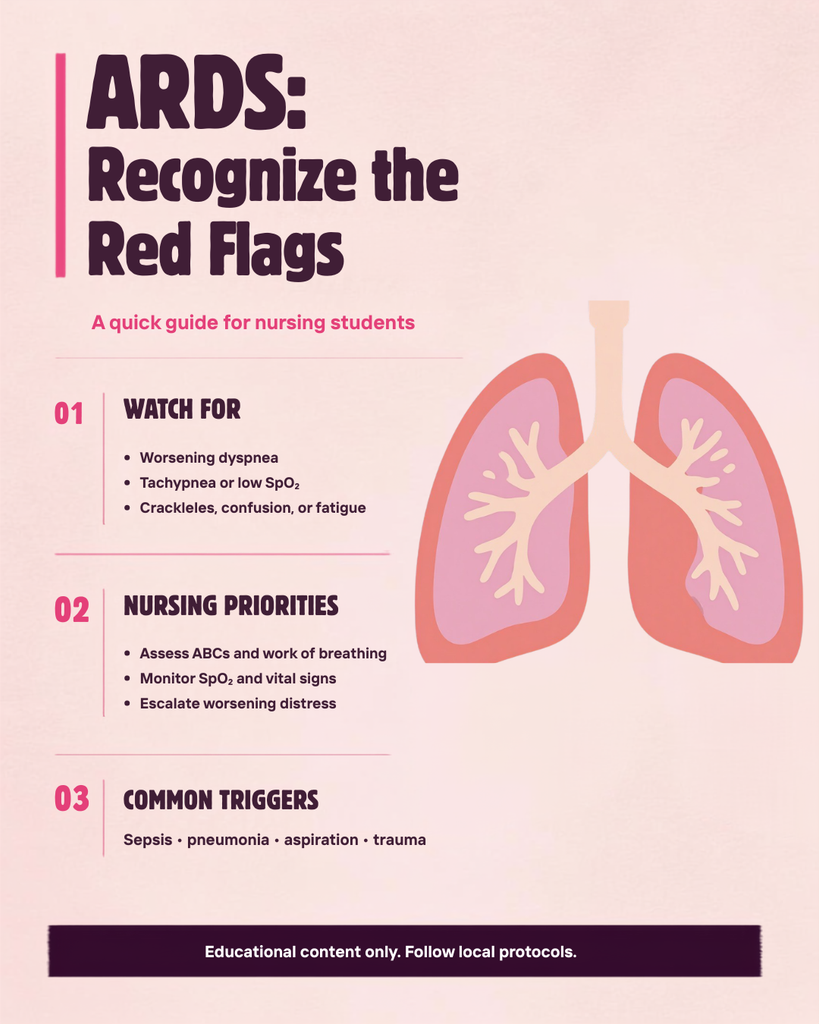
Common ARDS risk factors
- Pneumonia and other pulmonary infections
- Sepsis from a pulmonary or extrapulmonary source
- Aspiration of gastric contents
- Major trauma, pulmonary contusion, or inhalational injury
- Acute pancreatitis
- Massive transfusion or transfusion-related acute lung injury
- Near drowning and selected drug/toxin exposures
PE, stable COPD, obesity, isolated neuromuscular weakness, and isolated cardiogenic pulmonary edema can cause respiratory failure but are not, by themselves, defining ARDS insults. They may coexist with ARDS and complicate diagnosis.
Assessment and diagnostics
| Nursing/clinical assessment | Diagnostic evaluation |
|---|---|
| Work of breathing, respiratory rate/pattern, ability to speak, accessory-muscle use, breath sounds, cyanosis, mentation, hemodynamics, urine output, edema, and trends in oxygen requirement. | Pulse oximetry and ABG when indicated; chest radiograph/CT or lung ultrasound; ECG and laboratory evaluation; cultures and infection workup; echocardiography when hydrostatic edema or cardiac dysfunction is possible. |
| Review onset, aspiration, infection, trauma, transfusion, medications/toxins, comorbid heart/lung disease, fluid balance, and recent procedures. | Trend P/F or S/F ratio under documented support; evaluate alternative diagnoses such as cardiogenic edema, PE, diffuse alveolar hemorrhage, and acute exacerbation of interstitial lung disease. |
Management principles
Management combines treatment of the precipitating condition with supportive care that limits ventilator-induced lung injury. There is no single medication that reverses all ARDS.
Respiratory support
- Escalate supplemental oxygen and noninvasive support based on severity, work of breathing, mental status, hemodynamics, and response. Do not delay intubation when the patient is failing.
- For invasive ventilation, use lung-protective tidal volumes approximately 4–8 mL/kg predicted body weight and keep plateau pressure below 30 cm H₂O. Tidal volume is based on predicted—not actual—body weight.
- Use PEEP as part of an individualized strategy; higher PEEP may benefit selected moderate-to-severe ARDS but can cause overdistention or hemodynamic compromise.
- Use prone positioning for prolonged sessions (typically ≥12–16 hours/day) in severe ARDS and often in moderate-to-severe disease when indicated; use a trained team and pressure-injury/device-safety bundle.
- Consider neuromuscular blockade, inhaled pulmonary vasodilators as rescue, or venovenous ECMO only in selected patients under critical-care/specialty protocols. ECMO supports gas exchange; it does not treat the inciting cause.
Fluids and cause-directed care
- After shock is stabilized, a conservative fluid strategy is generally favored to reduce pulmonary edema while preserving organ perfusion.
- Treat the cause: timely antimicrobials/source control for infection, aspiration management, transfusion reaction management, trauma care, or other targeted therapy.
- Diuretics are not universal ARDS therapy; use when clinically indicated for fluid overload and perfusion/renal status permit. Thrombolytics treat selected unstable PE—not ARDS itself.
Nursing priorities
| Priority | Actions |
|---|---|
| Oxygenation/ventilation | Continuous SpO₂; trend ABGs when ordered; verify oxygen device, flow, FiO₂, ventilator settings, alarms, synchrony, ET tube depth, and secretion burden. |
| Perfusion | Trend MAP, rhythm, mental status, skin, urine output, lactate, fluid balance, and response to PEEP/position changes. |
| Prone safety | Secure airway/lines/drains; protect eyes and pressure points; use a checklist and trained team; reassess skin, tube position, and hemodynamics. |
| VAP/ICU prevention | Oral care, head-of-bed strategy when not prone/contraindicated, sedation and mobility protocols, DVT/stress-ulcer prophylaxis when indicated, nutrition, delirium and skin prevention. |
| Communication | Report increasing oxygen need, falling P/F or S/F ratio, rising PaCO₂ with acidemia, worsening work of breathing, ventilator dyssynchrony, new hypotension, oliguria, or altered mentation immediately. |
Avoid these common errors
- Calling all acute respiratory failure “ARDS.”
- Using ABG results alone to diagnose ARDS.
- Using actual body weight rather than predicted body weight for lung-protective tidal volume.
- Treating a single P/F ratio without documenting FiO₂, PEEP/support, trend, and clinical context.
- Assuming higher PEEP is always better; reassess compliance, oxygenation, blood pressure, and RV tolerance.
- Describing ECMO as definitive treatment rather than temporary organ support.
References
2. Grasselli G, et al. ESICM guidelines on acute respirahttps://pubmed.ncbi.nlm.nih.gov/37326646/tory distress syndrome: definition, phenotyping and respiratory support strategies. Intensive Care Med. 2023;49(7):727-759. doi:10.1007/s00134-023-07050-7.